connective tissue
Connective tissue
One of the four primary tissues of the body. It differs from the other three tissues in that the extracellular components (fibers and intercellular substances) are abundant. It cannot be sharply delimited from the blood, whose cells may give rise to connective tissue cells, and whose plasma components continually interchange with and augment the ground substance of connective tissue. Bone and cartilage are special kinds of connective tissue.
The functions of connective tissues are varied. They are largely responsible for the cohesion of the body as an organism, of organs as functioning units, and of tissues as structural systems. The connective tissues are essential for the protection of the body both in the elaborate defense mechanisms against infection and in repair from chemical or physical injuries. Nutrition of nearly all cells of the body and the removal of their waste products are both mediated through the connective tissues. Connective tissues are important in the development and growth of many structures. Constituting the major environment of most cells, they are probably the major contributor to the homeostatic mechanisms of the body so far as salts and water are concerned. They act as the great storehouse for the body of salts and minerals, as well as of fat. The connective tissues determine in most cases the pigmentation of the body. Finally, the skeletal system (cartilage and bones) plus other kinds of connective tissue (tendons, ligaments, fasciae, and others) make motion possible.
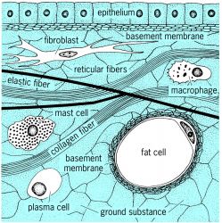
The connective tissues consist of cells and extracellular or intercellular substance (see illustration). The cells include many varieties, of which the following are the most important: fibroblasts, macrophages (histiocytes), mast cells, plasma cells, melanocytes, and fat cells. Most of the cells of the connective tissue are developmentally related even in the adult; for example, fibroblasts may be developed from histiocytes or from undifferentiated mesenchymal cells.
The extracellular components of connective tissues may be fibrillar or nonfibrillar. The fibrillar components are reticular fibers, collagenous fibers, and elastic fibers. The nonfibrillar component of connective tissues appears amorphous with the light microscope and is the matrix in which cells and fibers are embedded. It consists of two groups of substances: (1) those probably derived from secretory activity of connective tissue cells including mucoproteins, protein-polysaccharide complexes, tropocollagen, and antibodies; and (2) those probably derived from the blood plasma, including albumin, globulins, inorganic and organic anions and cations, and water. In addition, the ground substance contains metabolites derived from, or destined for, the blood.
All the manifold varieties of connective tissue may contain all the cells and fibers discussed above in addition to ground substance. They differ from each other in the relative occurrence of one or another cell type, in the relative proportions of cells and fibers, in the preponderance and arrangement of one or another fiber, and in the relative amount and chemical composition of ground substance. They are classified as:
1. Irregularly arranged connective tissue—which may be loose (subcutaneous connective tissue) or dense (dermis). The dominant fiber type is collagen.
2. Regularly arranged connective tissue—primarily collagenous—with the fibers arranged in certain patterns depending on whether they occur in tendons or as membranes (dura mater, capsules, fasciae, aponeuroses, or ligaments).
3. Mucous connective tissue—ground substance especially prominent (umbilical cord).
4. Elastic connective tissue—predominance of elastic fibers or bands (ligamentum nuchae) or lamellae (aorta).
5. Reticular connective tissue—fibers mostly reticular, moderately rich in ground substance, frequently numerous undifferentiated mesenchymal cells.
6. Adipose connective tissue—yellow or brown fat cells constituting chief cell type, reticular fibers most numerous.
7. Pigment tissue—melanocytes numerous.
8. Cartilage—cells exclusively of one type, derived from mesenchymal cells.
9. Bone—cells are predominantly osteocytes, but also include fibroblasts, mesenchymal cells, endothelial cells, and osteoclasts.
See Blood, Bone, Cartilage, Collagen, Histology, Ligament, Tendon
connective tissue
[kə′nek·tiv ′tish·ü]Connective Tissue
in animals, a tissue that develops from the mesenchyma and performs supportive, nutritive (trophic), and protective functions. A structural characteristic of connective tissue is the presence of well-developed intercellular structures (fibers and ground substance).
Connective tissue is classified as connective tissue proper, osseous tissue, and cartilaginous tissue, depending on the composition of the cells and the type, properties, and orientation of intercellular structures. Connective tissue proper may be irregularly or regularly arranged. Irregularly arranged connective tissue with fibers that are irregularly interwoven may be loose (areolar) or dense. An example of loose connective tissue is subcutaneous tissue, as well as connective tissue, which fills the spaces between organs and accompanies blood vessels. An example of dense connective tissue is the corium, or connective-tissue base of the skin. In regularly arranged connective tissue the fibers are arranged in certain patterns. This type of connective tissue occurs in tendons, fasciae, ligaments, and the sclera of the eye.
Reticular tissue and adipose tissue are two types of connective tissue having special properties. Another type of connective tissue is rich in pigment cells (for example, in the choroid of the eye). These tissues, together with blood and lymph, form the system of tissues within the body. Intercellular matter includes collagenous, elastic, and reticular fibers and the ground substance, which contains a large number of mucopolysaccharides. The fibers and ground substance are derived from fibroblasts—the principal cellular form of connective tissue.
Loose connective tissue also has macrophages, or histiocytes (cells that remove foreign particles and dead structures from tissue by phagocytosis), mast cells (cells that contain heparin, histamine, and other biologically active substances), fat, pigment, and plasma cells, and various types of blood leukocytes. Loose connective tissue, which fills the spaces between organs, vessels, nerves, muscles, and other body structures, forms the internal medium through which nutrients are delivered to cells and the products of metabolism are removed. Its extensive distribution and role in cell nutrition and protective processes make this tissue a participant in almost all of an animal’s physiological and pathological reactions, including inflammation, physiological and reparative regeneration, healing of wounds, and sclerotic processes.
Connective tissue with marked protective functions is characterized by a relatively large number and diversity of cells, including blood leukocytes. In predominantly supportive connective tissue, intercellular structures predominate and cells are represented only by fibroblasts or their analogues, including cartilage and bone cells.
REFERENCES
Eliseev, V. G. Soedinitel’naia tkan’. Moscow, 1961.Khrushchov, N. G. Funktsional’naia tsitokhimiia rykhloi soedinitel’noi tkani. Moscow, 1969.
Khrushchov, N. G. Gislogenez soedinitel’noi tkani. Moscow, 1976.
N. G. KHRUSHCHOV
