Encyclopedia

Mesoderm

Also found in: Dictionary, Medical, Wikipedia.

mesoderm

[′mez·ə‚dərm]
(embryology)
The third germ layer, lying between the ectoderm and endoderm; gives rise to the connective tissues, muscles, urogenital system, vascular system, and the epithelial lining of the coelom. Also known as mesoblastema.
McGraw-Hill Dictionary of Scientific & Technical Terms, 6E, Copyright © 2003 by The McGraw-Hill Companies, Inc.
The following article is from The Great Soviet Encyclopedia (1979). It might be outdated or ideologically biased.

Mesoderm


mesoblast, in multicellular animals (except sponges and coelenterates), including man, the middle germ layer, found between the ectoderm (the outer germ layer) and the endoderm (the inner) as a result of gastrulation.

In protostomes (most invertebrates), the mesoderm is formed from teloblasts—large cells lying between the ectoderm and endoderm at the posterior end of the embryo that during gastrulation enter the primary body cavity, where they multiply and develop into two mesodermal bands. In most deuterostomes (echinoderms, brachiopods, chaetognaths, acranians, cyclostomes, fish, amphibians), the mesoderm is formed enterocoelically—that is, from separating portions of the wall of the primitive gut, or enteron. In other deuterostomes (reptiles, birds, and mammals), because of secondary changes in the process of germlayer individuation, the mesodermal rudiment becomes part of the primitive ectodermal layer during the blastula stage and later becomes a third germ layer, the mesoderm.

Figure 1. Diagram of mesodermal development in annelids: (1), (2), (3) successive stages; (a) ectoderm, (b) endoderm, (c) mesodermal band, (d) somite, (e) coelom, (f) dorsal mesentery, (g) musculature, (h) gut, (i) ventral mesentery, (j) ventral nerve trunks, (k) inner wall of coelom

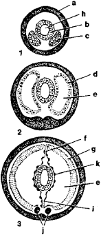
In flatworms and nemertines, the mesodermal bands give rise to the connective tissue that fills the spaces between the viscera. In annelids and arthropods, they divide into paired somites with a secondary body cavity, or coelom (see Figure 1). The longitudinal muscles of the body and the excretory organs develop from the walls of the coelom. The mesoderm develops more or less similarly in the various groups of vertebrates (see Figure 2).

Figure 2. Diagram of development of organs from mesoderm in a higher vertebrate (cross section of embryo): (a) neural tube, (b) dermatome, (c) ectoderm, (d) myotome, (e) sclerotome, (f) nephrotome, (g) outer layer of splanchnotome, (h) endoderm, (i) inner layer of splanchnotome, (j) aortal endothelium, (k) coelom, (I) notochord

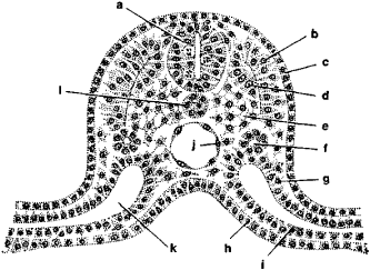
The rudimentary notochord develops in the dorsal part of the embryo. The mesoderm divides into metameric somites on both sides of the notochord. These somites are initially connected to nonsegmented ventral portions of the mesoderm, called lateral plates (splanchnotomes), by narrow segmented stalks, or nephrotomes. The wall of each somite then differentiates into sclerotomes, dermatomes, and myotomes. The sclerotomes form the axial skeleton and connective tissue; the dermatomes, the connective-tissue layer of the skin; and the myotomes, the skeletal musculature. The nephrotomes differentiate into the renal tubules of the primitive kidney (and later, in higher vertebrates, of the secondary kidney), and the ducts of the urogenital system.

The splanchnotomes divide into two layers, inner (visceral) and outer (parietal), and the coelom forms between them. The visceral layer adjoins the endoderm and gives rise to the smooth musculature of the intestine, to the blood vessels and blood cells, and to the lining of the body cavity. The parietal layer adjoins the integuments and lines the coelom. The genital ridges, rudiments of the gonads, arise in the epithelium of the splanchnotomes. The right and left lateral plates grow together over the intestine to form the mesentery.

REFERENCES

Davydov, K. N. Kurs embriologii bespozvonochnykh. Petrograd-Kiev, 1914.
Ivanov, P. P. Obshchaia i sravniteVnaia embriologiia. Moscow-Leningrad, 1937.
Shmal’gauzen, I.I. Osnovy sravniteVnoi anatomii, 4th ed. Moscow, 1947.
Shmidt, G. A. Embriologiia zhivotnykh, 2 parts. Moscow, 1951–53.
Tokin, B. P. Obshchaia embriologiia. Moscow, 1970.

T. A. DETLAF and A. V. IVANOV

The Great Soviet Encyclopedia, 3rd Edition (1970-1979). © 2010 The Gale Group, Inc. All rights reserved.
Mentioned in
References in periodicals archive
Insights from diploblasts: the evolution of mesoderm and muscle.
In B, histology of the embryo with 9 days, evidencing the neural tube (Nt), the intermediate mesoderm (MI) and the beginning of the heart formation (h).
Biased cell rotation is seen as a dominant CW bias in day 5 cardiac mesoderm and CCW biases in day 3 endoderm, day 7 mid/hindgut, and day 5 neural induced stages.
(See figure) In placing the embryo outline over the iris chart, we see the areas of the ectoderm, the mesoderm, and the endoderm relate to the location of the various organs and body structures.
Also, in the Mixl1-null mouse embryos can be confirmed the independence of migratory movements of embryonic endoderm and mesoderm [91], suggesting that the establishment of the two lineages could be originated before the hypoblast displacement [88, 90, 91].
The first group arises as a result of developmental failure of a segment of the mesoderm and comprises three of the defects, which include diaphragmatic defect (which results from total or partial failure of the transverse septum to develop), pericardial defect (which is closely related to faulty development of the transverse septum), and intracardiac lesions (which is the result of faulty development of the epimyocardium, which is derived from the splanchnic mesoderm) [8, 22, 24].
An aqua lifting automatic injector, EPN can be used for therapy to rejuvenate the skin using its micro needles to stimulate the mesoderm without leaving any scars and boosts the skin's natural healing process and the collagen rearranging process.
These particular stem cells have been detached from the sub-amnion to the perivascular region of the umbilical cord, with its origin being extra-embryonic or embryonic mesoderm.1 WJMSCs differentiate into cardiomyocytes and combine with the ischaemic cardiac tissues which helps to improve cardiac function.2
At the limb initiation stage, limb buds are formed from the lateral plate mesoderm while apical ectoderm ridge (AER) is formed by early limb mesoderm growth1.
During the embryonic stage of development, limbs arise as a condensation of cells from lateral plate mesoderm and its ectodermal covering (Al-Qattan et al., 1998).
MSCs are cells with high in vitro self renewal capacity and ability to differentiate into multiple mesoderm ectoderm and endoderm lineages.
Copyright © 2003-2025 Farlex, Inc Disclaimer
All content on this website, including dictionary, thesaurus, literature, geography, and other reference data is for informational purposes only. This information should not be considered complete, up to date, and is not intended to be used in place of a visit, consultation, or advice of a legal, medical, or any other professional.